
New Images Reveal the Ravaged Landscape of COVID-19 Lungs in Unprecedented Detail
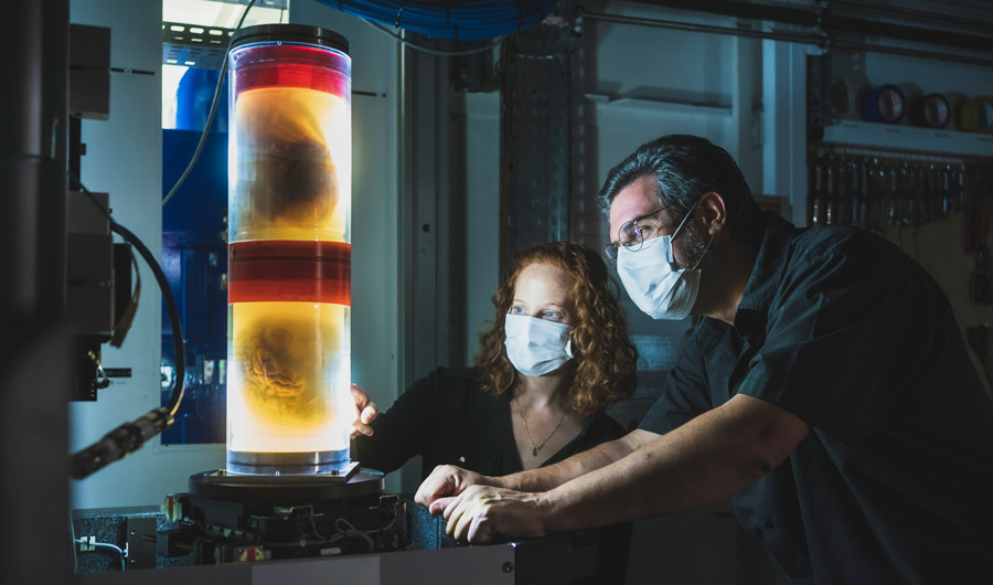
Claire Walsh and Paul Tafforeau at the European Synchrotron Radiation Facility (ESRF) with two samples prepared for HiP-CT imaging.
ESRF/Stef Candé
Used with permission.
(Inside Science) -- A revolutionary tool designed to broaden our understanding of human anatomy has for the first time provided scientists with a cellular-level look at lungs damaged by COVID-19.
In healthy lungs, the blood vessel system that oxygenates the blood is separate from the system that feeds the lung tissue itself.
But in some severe respiratory illnesses, such as pneumonia, pressures caused by the infection can lead blood vessels in the heart and lungs to expand and grow, sometimes cutting through the body and forming channels between parts of the pulmonary system that shouldn't be connected. Similarly, COVID-19 infections can create the same types of abnormal channels. The channels give unoxygenated blood coming into the lungs an alternate exit ramp, allowing it to essentially skip the line and shoot back into the body without picking up any oxygen molecules first. Scientists believed that this could be a cause of the low blood oxygen levels sometimes experienced by COVID-19 patients, a condition known as hypoxemia.
"Blood vessel growth is a very controlled process," said Claire Walsh, a medical engineer at University College London and the first author of the imaging study, published in the journal Nature Methods. "It should be in this lovely tree-like branching structure. And you look at the COVID lungs, and you can just see it's in these big clumps of really dense vessels all over the place, so that it just looks … wrong."
Walsh's team, which included clinicians from Germany and France, has procured sharper-than-ever images of these warped structures, thanks to an imaging technique known as HiP-CT, or Hierarchical Phase-Contrast Tomography, which allows them to zoom in on any body part with 100 times the resolution of a traditional CT scan. Although the technique can only be used to capture images of samples removed from a body and preserved in a way that minimizes interference (rather than of organs that are still part of a living person), in pairing it with the world's brightest X-rays at the European Synchrotron particle accelerator, the researchers hope to build a visual database of not only lungs infected with COVID-19, but other, healthy organs throughout the body.
"It gives you a view of human anatomy that so many people have never seen before," Walsh said of the tool.
The Human Organ Atlas, the database that the international team is currently putting together, will include HiP-CT scans of everything from brains to kidneys. There's a good chance that capturing snapshots of how different diseases operate will be a boon to researchers working on treatments.
Walsh herself has high hopes for what the technique can teach us about all sorts of pulmonary conditions. In the lungs, "to understand what's happening to the organ as a whole, it's really important that you understand the overall architecture of the main branching trees,” she said. “Just looking at a really small bit in isolation, it can be really hard to understand what's going on in the organ. It's so interconnected, and I think that makes the lungs particularly good for this technique."