Operating Cells Via Joystick

(Inside Science) -- Biomedical research could someday look a lot like playing video games thanks to a new device that allows users to manipulate cells with the swerve of a joystick.
A team of physicists and engineers at Ohio State University in Columbus, Ohio developed the device from a tiny piece of square-centimeter silicon inlaid with rows of zigzagging magnetic wires. At each corner, the wire behaves like two magnets pointed north to north or south to south. The fields of the two magnets create a point of strong attraction just above them. A nearby magnetic object, such as a magnetically-tagged cell is attracted to the corner and gets stuck there
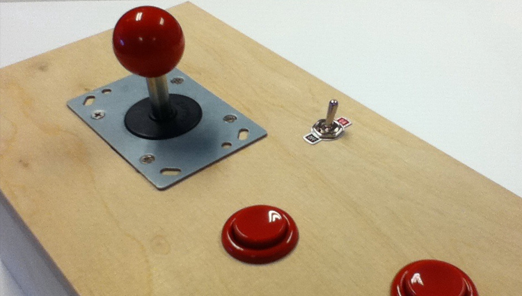
To get the particles moving, the researchers then place two magnetic fields around the chip one in the plane of the chip and the other perpendicular to it. By flipping the direction of these fields, the researchers can guide tagged cells along the zigzagging wire and even make them jump from one wire to the next. The researchers computerized the magnetic field switching so that a user steered the cells by simply handling a joystick.
The team at OSU put the device through its paces with magnetically-tagged T-cells, the body's guardians against infection. They snapped the cells to attention at one end of the chip, marched them down to the other end, and made them hop from one wire to another, reaching speeds of about 20 micron, or about a one-fifth the width of a human hair, per second.
Jeffrey Chalmers, the chemical engineer who tagged the T-cells for the experiment, said that the device would be ideal for examining tumor cells. To study biopsied tumors, researchers often treat them with enzymes, which break them down into their constituent cells. Researchers then separate cancerous cells they want to study from healthy cells like fat and blood.
"Part of the problem with cancer ... is that it's our own cells going haywire, so it's a heck of a lot harder to figure out what's different," Chalmers said. With this method, he said, researchers could magnetically tag the well-understood healthy cells and then remove them from a sample, leaving only the cancerous cells. Chalmers said this would be a boon to both a researcher studying a specific type of cancer or a clinician diagnosing a patient.
"The technology to do high-level analysis is pretty amazing, but it's only as good as the purity of the sample you start with," Chalmers said. "The more you can separate them out, [the more] you know what you're looking at."
The small magnetic fields are gentle on specimens; the device works on a flat surface, an improvement over other methods; and it's also cost-effective. The project's principal investigator, physics professor Ratnasingham Sooryakumar, said that the whole set up only costs about $200. He said it could easily be scaled up to a square centimeter silicon platform, with about 10,000 tiny traps, or scaled down to manipulate organelles within a single cell.
Sooryakumar said that scaling up would lead to a "lab on a chip," where researchers could cheaply and easily look at distinctive behavior within large populations of cells, making it easier to draw firm conclusions.
"You can look at each cell rather than averaging it out, and say, 'the cell on vertex number 348 did this,'" Sooryakumar said. "When you actually have 10,000 of them to analyze the data, you can understand stat distributions that we normally would not have gotten in ensemble measurements, and that's a huge thing."
Sooryakumar envisions embedding the device into containers that hold tiny amounts of fluid, like blood. By tagging a certain kind of particle, researchers could begin separating, say, viruses from healthy blood cells. Chalmers added it could be used to study cancer in blood samples.
"One in a million or one in a billion cells in your blood could be cancer," Chalmers said, but the technique could achieve higher concentrations of cancer cells to study by tagging and removing healthy blood cells.
Prem Thapa, a researcher at Kansas State University in Manhattan, Kan., who was not involved in the study, called the approach "interesting and innovative," adding that the technique had advantages over existing optical manipulation methods.
"The significance of these studies is high," Thapa said. But he pointed out that electrically excitable neurons or muscle cells may not take so kindly to magnetic manipulation.
Thapa's K-State colleague, physicist Brett Flanders, was impressed by the results but called the demonstration "simple."
"As with ... all potential biophysical applications, there's a lot more work to do," said Flanders said. "I'm looking forward to seeing what comes next."
