
Eye Damage In Diabetes Could Stem From The Cells Used To See
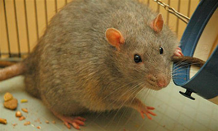
A Zucker rat. This rat has a genetic disorder that causes obesity, and has developed diabetes as a result.
Joanna Servaes via wikipedia commons | http://bit.ly/15ZmjMr
(ISNS) -- A potentially blinding condition may have its cause in the very cells that mediate vision, a new study suggests. Ophthalmology researchers have implicated photoreceptors, the cells that sense light and are the first step in sending visual signals to the brain, in the production of compounds that can cause both inflammation and new blood vessels to invade the retina. This retinopathy, a common complication in diabetes, can impact vision through swelling of the retina, leaky capillaries, and death of cells in the eye.
Because a hallmark of diabetic retinopathy is neovascularization – the rapid growth of abnormal blood vessels in the eye – either white blood cells or the endothelial cells that make up these vessels were thought of as culprits. But, said Timothy Kern, a professor of medicine at Case Western Reserve University, in Cleveland, these cells are “victims, rather than mediators,” of toxic compounds produced by another cell type. Kern and colleagues have now identified that in the diabetic eye, photoreceptors are a source of tissue-damaging free radicals, which kick off a cascade of inflammation and blood vessel growth that can lead to vision loss. Their research was recently published in the Proceedings of the National Academy of Sciences.
Using diabetic mice, the scientists first identified where in the retina – the “seeing” layer of the eye – certain free radical compounds were present. Fluorescent staining of cells under a microscope showed that they were concentrated around the photoreceptors. These cells – rods and cones – are more metabolically active at night, when they are both sensitized to pick up any light in the darkness, and are also undergoing cellular regeneration. Kern and his team observed that more free radicals were also generated at night. Stopping photoreceptors from experiencing complete darkness at night is a new therapeutic angle that is being investigated, said Kern. “In the future it may be possible to use lights to inhibit retinopathy by keeping the photoreceptors from going into the dark when they become most active.”
Human trials using dim illumination of one eye during the night have shown promise in reducing diabetes-related retinal symptoms.
To definitively determine that photoreceptors were in fact generating the free radicals, Kern and his team studied mice that had no photoreceptors, either because of a genetic mutation or an experimental manipulation that killed the cells. These blind mice had lower levels of oxidative compounds, consistent with the idea that the photoreceptors were the major source. They also had reduced amounts of inflammatory proteins in the retina, which are normally produced in response to oxidative stress.
Previous research has found that diabetic animals without photoreceptors also have less retinal capillary damage, which is an early sign of diabetic retinopathy. When capillaries die, the retina doesn’t get enough oxygen, and starved cells will send out signals to get new blood vessels to grow, which can lead to vision problems.
“We’ve identified the mechanism by which photoreceptors could be causing this capillary degeneration,” said Kern, “but there’s a huge black box between the early and end points that we’re trying to resolve.”
Part of the problem may be that photoreceptors are naturally very metabolically active cells, since they mediate vision, and therefore contain a lot of mitochondria, which generate cellular energy. Kern says that in diabetes, mitochondria can be abnormal in other organs like the heart and kidney, so there may be reason to think mitochondria in the eye are contributing to retinopathy.
Bruce Berkowitz, who researches diabetic retinopathy at Wayne State University, describes the photoreceptors as working overtime in the diabetic eye. “This paper presents something that was not really suspected, which is that the photoreceptors are possible disease generators,” said Berkowitz, who was not a part of the study. “How and why this is happening is a whole new field of research.”
The next step, said Kern, is an ongoing investigation of a chemical that could shut off the mitochondria’s ability to make certain free radicals, which would slow the process that leads to retinopathy without killing photoreceptors.
Specific targeting of photoreceptors with antioxidants, which can be obtained through the diet, might be worth investigating, added Berkowitz. “There are lots of diabetic Americans who are losing their sight every day, and that is a reason to pursue this work.”
Amanda Alvarez has written about science for the Milwaukee Journal Sentinel, Yale Medicine, and GigaOM. She received her PhD in Vision Science from the University of California, Berkeley, and tweets at @sci3a.